INTRODUCTION
In computed tomography (CT), radiation is used to obtain images. These images are reconstructed using computer-analysis, providing detailed cross-sectional imaging.
Computed tomography dramatically increased diagnostic accuracy of imaging studies. In addition to diagnosis, computed tomography offers the possibility to performed important therapeutic interventions using a minimally invasive approach (such as image-guided percutaneous drainage of intraabdominal abscesses).
During the last two decades, the use of computed tomography has dramatically increased.
CONCERNS
This dramatic increase of the use of computed tomography raised concerns about the potential problems and associated patient harm. Potential complications include the side-effects of the ionizing radiation, specifically regarding carcinogenesis.
COMPUTED TOMOGRAPHY – CARCINOGENETIC EFFECTS OF RADIATION
The carcinogenetic effects of radiation are well-known. It has been estimated that computed tomography and nuclear medicine procedures are the main source of exposure of the general population to radiation. About 25 % of the exposure of the US population to radiation is due to these two imaging modalities (data from 1997 – 2007, the percentage could be even higher today).
Radiation doses during computed tomography ranges widely, depending on many factors. Diagnostic computed tomography exposes to a radiation dose ranging from 1 to 15 mSv (millisieverts), during a simple procedure. This dose is comparable to the low-radiation dose received by atomic bomb survivors (5 – 20 mSv). These survivors were presented later with a significantly increased incidence of several types of cancers, including thyroid cancer. These cancers were developed many years after the exposure to radiation. Increased exposure to ionizing radiation also occurs in workers in the nucleal industry; these people are exposed to an average dose of 20 mSv. This dose is equivalent to one or two CT scans.
The risk of cancer risk following exposure to radiation during CT may appear small on an individual basis; however, the overuse of computed tomography may be associated with a real public health issue. In studies using mathematical prediction models, it has been estimated that about 1.5 – 2 % of future cancers in the US might be caused by computed tomography. This is an important consideration, since about 29.000 new cases and 15.000 deaths per year will be attributed to radiation exposure during CT examinations. This risk remains increased in the long term (latent period even more than 10 years).
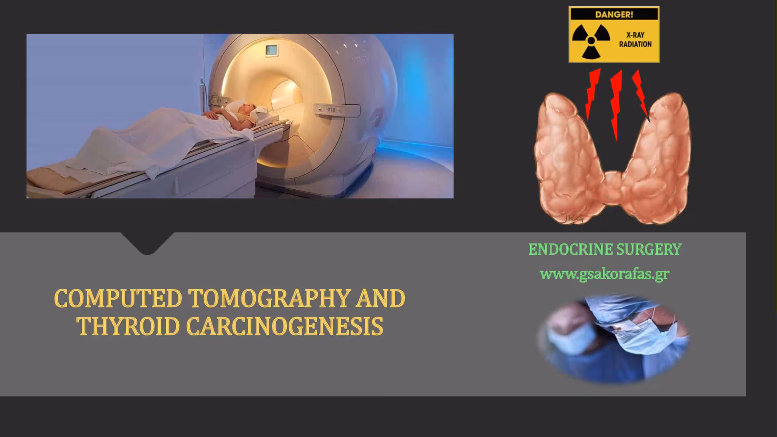
COMPUTED TOMOGRAPHY AND THYROID CARCINOGENESIS
Thyroid parenchyma is highly sensitive to the carcinogenetic effects of ionizing radiation. This is especially true in children. The risk of thyroid cancer development following CT is significantly increased not only in children, but also in adults, especially in women and patients aged < 45 yrs. A strong “dose-risk” relationship exists. The risk also correlates with the anatomical region scanned and is highest in patients undergoing CT of the neck.
CONCLUSIONS
Thyroid cancer development is a potential long-term complication of the exposure to ionizing radiation during CT. CT is a highly valuable imaging method; however, it should be used judiciously. Appropriate use of CT and reduction of radiation dose per examination may optimize the benefit-risk ratio of computed tomography.
JNCI Cancer Spectrum 2020